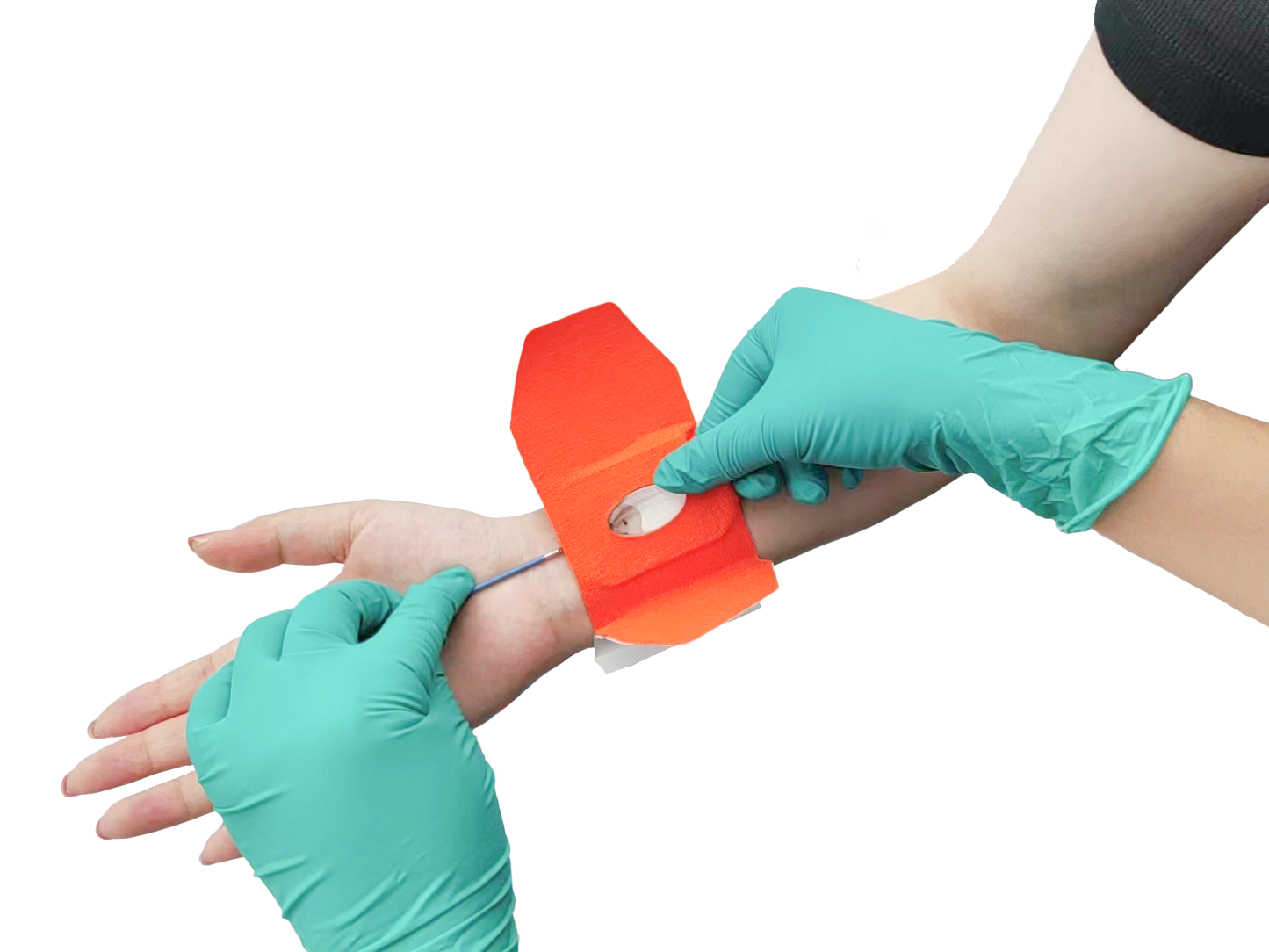
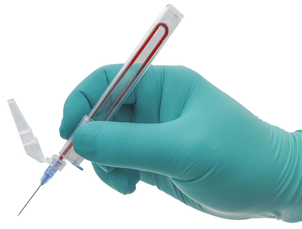
FEMO-SUPERBOWL™ Femoral Hemostasis Compressor
One compressor for every femoral closure — from routine 6F angiography to ECMO decannulation. Focal spherical compression that preserves distal flow and leaves no foreign material behind.
Point Compression That Keeps Blood Flowing
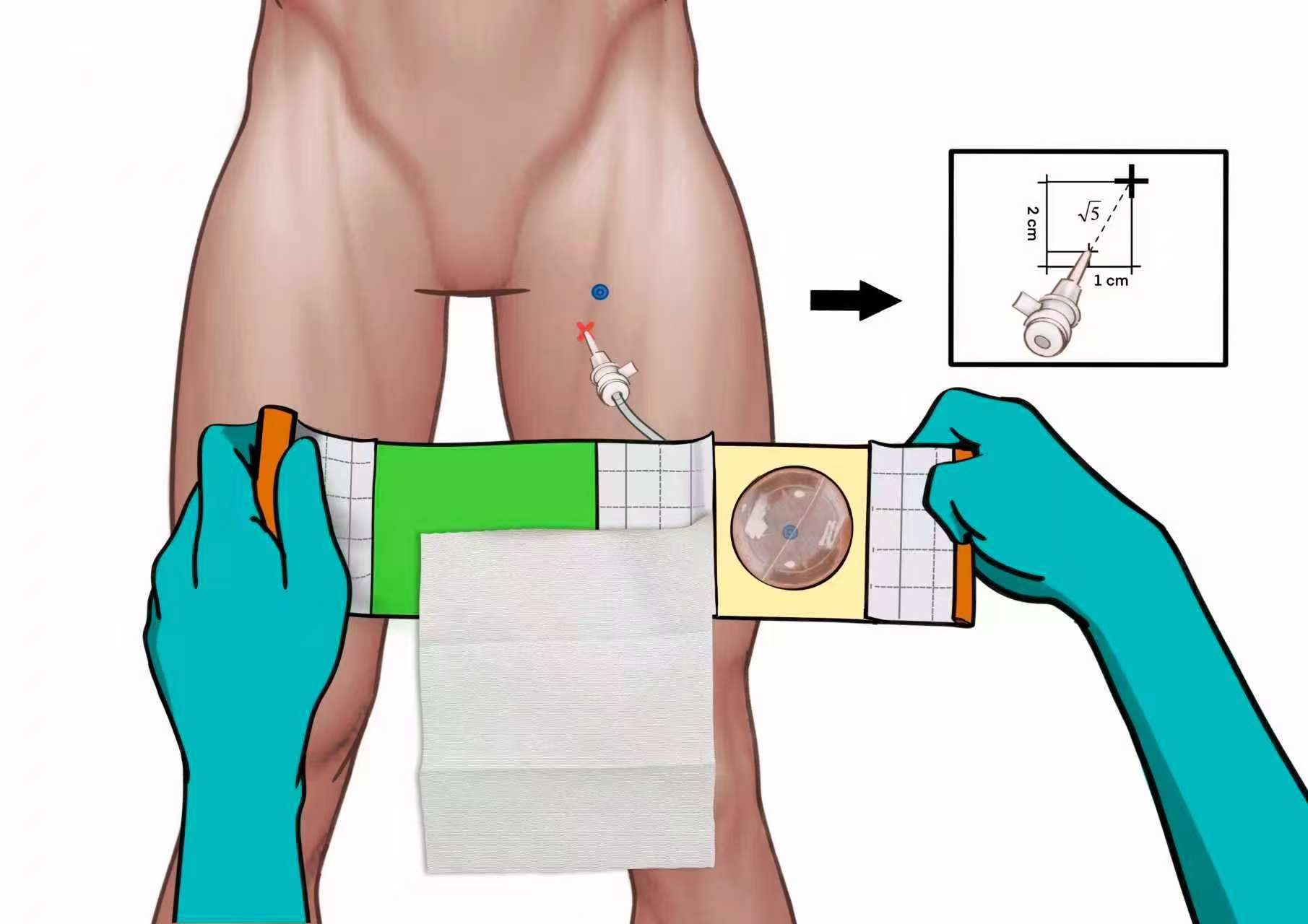
The rigid yet elastic spherical bowl transmits focal pressure through adipose and muscle layers directly to the femoral arteriotomy. Unlike flat plates or inflatable designs that compress the entire thigh, the bowl curvature concentrates force exactly where it is needed — achieving hemostasis without occluding collateral vessels.
The outer reference ring is engineered to match the anatomical skin-to-artery offset of √5 cm. Aligned with the skin puncture site along the femoral axis, the bowl center sits directly over the subcutaneous arterial puncture — no guesswork, no fluoroscopy. The transparent bowl then allows continuous visual monitoring of the puncture site throughout the compression period.
- Compatibility5F–23F sheaths, from diagnostic angiography to ECMO decannulation
- OrderingREF 216880 · Sterile · 1 unit per pouch
- PerfusionDistal flow typically restored within about 60 seconds of application
- EfficiencyCompression time reduced by half or more versus traditional manual protocols
Engineered for Femoral Hemostasis
Spherical Bowl, Deep Point Compression
A sphere focuses force to a single point. The bowl compresses the deep arterial defect while surrounding tissue stays under far less pressure — hemostasis without occluding collateral vessels.
√5 cm Reference Ring
The ring matches the anatomical offset from skin to artery. Aligned with the skin puncture site along the femoral axis, it places the bowl center directly over the subcutaneous arterial puncture.
Transparent Visual Window
Continuous visual confirmation of the puncture site throughout compression. Clinicians can monitor for bleeding or hematoma without disturbing the device.
Flow-Preserving Fixation
The elastic tape system secures the device without a tourniquet effect, preserving profunda femoris and superficial femoral artery flow and minimizing thrombosis and occlusion risk.
No Foreign Material
Pure external compression — no intraluminal implant and no vessel wall injury. The access site is fully preserved for future procedures.
One Operator, One Device
Rapid, reproducible bedside application with minimal bruising — designed for daily intervention and proven for extreme cases.
Measured at the Bedside
Average time to return of normal rSO₂ and pedal pulse after device application — confirming flow is maintained, not occluded.
Preserved distal flow minimizes thrombosis and occlusion, keeping the vessel ready for future interventions.
Compression duration reduced by half or more compared with traditional manual protocols — tailored to sheath size.
Proven Across the Femoral Access Spectrum
Standard & Complex Interventions
- Diagnostic angiography (5F–6F)
- Peripheral vascular interventions (6F–8F)
- Transcatheter aortic valve replacement — TAVR (8F–14F)
- Endovascular aneurysm repair — EVAR
- Impella / mechanical circulatory support
VA-ECMO Decannulation (15F–19F)
ECMO arterial cannulas represent the most demanding hemostasis challenge. FEMO-SUPERBOWL™ enables:
- 10–20 minutes of manual compression after decannulation, then device application for 12–24 hours
- No need for surgical cutdown or suture-based closure devices
- Reduced ICU bed rest and faster liberation from immobility
FEMO-SUPERBOWL™ vs. Invasive Closure Devices
Suture-Based Closure
- Leaves permanent foreign material in the vessel
- Risk of infection, suture entanglement and vessel stenosis
- Failure rate up to 10%
Plug-Based Closure
- Retained collagen or polymer plug
- Risk of embolization, pseudoaneurysm and late infection
- Not suitable for small or calcified vessels
FEMO-SUPERBOWL™
- External focal compression — no foreign material, no vessel wall injury
- Preserves the access site for future procedures
- Cost-effective, simple application
Eight Steps to Reproducible Hemostasis
Prepare Components
Remove the sterile packaging and release the tape securing clip. Remember: the visible skin entry point is vertically offset from the actual subcutaneous arterial puncture by approximately √5 cm, so the spherical center must be aligned over the arterial puncture site.
During Catheter Removal
Apply digital pressure primarily targeting the subcutaneous arterial puncture site before withdrawing the catheter. Maintain the pressure and gradually increase compression force while slowly withdrawing.
Immediately Post-Removal
Continue firm, uninterrupted digital compression over both the arterial puncture and the skin entry point. Place a sterile gauze pad over the entry point and maintain adequate pressure for 5 minutes — or 10–20 minutes before device application for large-bore catheters (8F–23F, ECMO cannulas).
Device Positioning
Align the center of the hemispherical surface directly over the subcutaneous arterial puncture site. Ensure precise alignment throughout positioning — do not release pressure during placement or adjustment.
Secure the Short Tape
Confirm hemostasis at the puncture site. Stretch the shorter tape on the right side of the sphere to automatically detach the release liner, then adhere it to the outer thigh while maintaining manual pressure.
Secure the Long Tape
Wrap the longer tape around the patient thigh. Adhere it to the inner thigh first, then crosswise to the abdomen, ensuring the tape presses firmly against the sphere to maintain balance.
Final Adjustments
Adjust tape tension to stabilize the sphere and prevent displacement. Ensure the visual window on the sphere remains unobstructed for monitoring the puncture site.
Reinforce if Needed
For insufficient pressure, sphere misalignment or obese patients, use backup tape to reinforce fixation — applied diagonally or circumferentially based on clinical judgment.
Compression Duration Guidelines
| Sheath size | Typical compression duration | Procedure type |
|---|---|---|
| 5F–8F | 4–6 hours | Standard femoral interventions |
| 8F–12F | 6–8 hours | Larger sheath procedures (TAVR, EVAR) |
| ≥15F | Up to 24 hours | ECMO cannulas and large-bore access |
Clinical note: compression duration should be adjusted based on intraprocedural anticoagulation, patient coagulation status, procedural complexity and sheath size. The times above are general guidelines — clinical judgment should always prevail. Once hemostasis is confirmed, remove the system.
Product Specifications
| Catalog number | Product name | Sheath size compatibility | Sterility | Quantity |
|---|---|---|---|---|
| 216880 | FEMO-SUPERBOWL™ Arterial Hemostasis Compressor | 5F–23F (diagnostic angiography to ECMO) | Sterile | 1 unit / pouch |
Request Pricing, Samples or Distribution Terms
Contact our team for FEMO-SUPERBOWL™ product information, pricing, samples or distribution enquiries. We reply within one business day.
Related Products